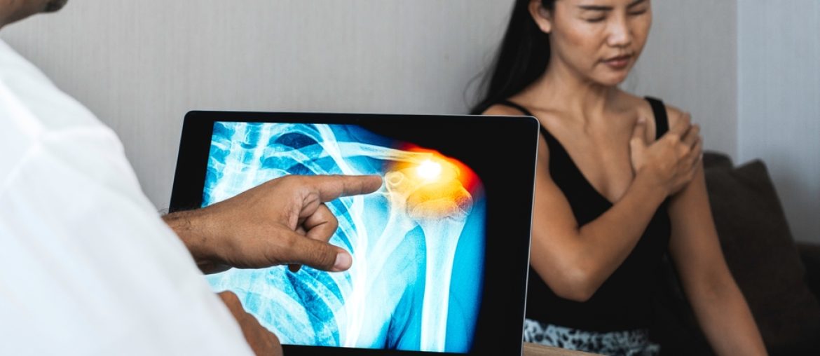
Accurate diagnosis is crucial in orthopedic care, where precise understanding of musculoskeletal issues can make the difference between a speedy recovery and prolonged discomfort. At MD West One in Omaha, NE, Dr. Michael A. Del Core utilizes a variety of advanced imaging techniques to diagnose orthopedic conditions and determine the most effective treatment plans. Among the most commonly used diagnostic tools in orthopedics are X-rays, MRI scans, and CT scans. Each of these imaging methods serves a unique purpose and provides critical insights into different types of injuries and conditions affecting the bones, joints, and surrounding tissues.
X-Rays: The Foundation of Orthopedic Imaging
X-rays are one of the most widely used diagnostic tools in orthopedics. They are a type of electromagnetic radiation that can pass through the body, allowing for the visualization of bones and certain tissues. X-rays are particularly effective in diagnosing fractures, dislocations, and joint conditions such as arthritis.
- How It Works: During an X-ray procedure, a machine sends a beam of X-rays through the body, which is absorbed differently by various tissues. Dense structures, like bones, absorb more X-rays and appear white on the resulting image, while softer tissues allow the X-rays to pass through, appearing darker.
- Common Uses in Orthopedics: X-rays are most commonly used to identify bone fractures, dislocations, bone deformities, and joint degeneration. They can also help detect infections, tumors, and other abnormalities in the bones.
- Limitations: While X-rays are excellent for imaging bones, they are less effective for soft tissues such as muscles, tendons, and ligaments. For these structures, more advanced imaging techniques like MRI or CT scans are often required.
- Safety: X-rays expose the body to a small amount of radiation, but this is generally considered safe, especially when used occasionally. Protective measures, such as lead aprons, are used to minimize exposure to sensitive areas.
At MD West One, X-rays are often the first imaging tool used to assess injuries or conditions affecting the bones and joints. They provide quick and valuable insights that help guide further diagnostic or treatment decisions.
MRI: Uncovering Soft Tissue and Complex Injuries
Magnetic Resonance Imaging (MRI) is a powerful diagnostic tool that provides detailed images of soft tissues, including muscles, tendons, ligaments, and cartilage, as well as bones. Unlike X-rays, which rely on radiation, MRI uses magnetic fields and radio waves to produce highly detailed cross-sectional images of the body.
- How It Works: An MRI machine creates a strong magnetic field that aligns the hydrogen atoms in the body. When radio waves are sent through the body, these atoms produce signals that are captured by the machine and translated into detailed images. The result is a series of high-resolution images that show the internal structures of the body in great detail.
- Common Uses in Orthopedics: MRI is particularly useful for diagnosing soft tissue injuries such as ligament tears, muscle strains, tendonitis, and herniated discs. It is also commonly used to assess joint conditions like meniscus tears in the knee or rotator cuff injuries in the shoulder. Additionally, MRI is often employed to detect bone tumors, infections, and spinal cord abnormalities.
- Advantages: MRI provides a clear and comprehensive view of soft tissue structures, which are often difficult to visualize with other imaging methods. This makes it an invaluable tool for diagnosing complex injuries that involve both bones and soft tissues.
- Limitations: While MRI is extremely effective, it is also more time-consuming and expensive compared to other imaging techniques. Additionally, patients with certain metal implants, such as pacemakers, may not be able to undergo an MRI due to the strong magnetic field.
For patients at MD West One experiencing soft tissue injuries or complex musculoskeletal conditions, MRI scans are often recommended to get a complete picture of the issue and to develop an accurate and effective treatment plan.
CT Scans: Detailed Views of Bone and Joint Structure
Computed Tomography (CT) scans are another essential diagnostic tool in orthopedics, providing highly detailed images of the bones and joints. A CT scan combines X-ray technology with computer processing to create cross-sectional images of the body, offering more detail than a standard X-ray.
- How It Works: A CT scan involves taking multiple X-ray images from different angles around the body. A computer then compiles these images into a single, comprehensive picture, allowing for detailed views of both bones and surrounding tissues.
- Common Uses in Orthopedics: CT scans are particularly valuable for diagnosing fractures that are difficult to see on regular X-rays, such as stress fractures or fractures that occur in complex areas like the spine, pelvis, or wrists. They are also useful for visualizing joint problems, including cartilage damage and bone spurs.
- Advantages: CT scans provide a more detailed view of bone structure than traditional X-rays, making them ideal for diagnosing complex fractures or bone deformities. They also offer faster imaging times compared to MRI, making them a good option when time is a critical factor.
- Limitations: CT scans expose patients to more radiation than X-rays, although the dose is still considered safe for most patients. Additionally, while CT scans provide excellent images of bones, they are less effective for soft tissue visualization compared to MRI.
At MD West One, CT scans are frequently used when detailed imaging of bone injuries is required, especially in cases where traditional X-rays do not provide sufficient information. This advanced imaging tool helps ensure that fractures and other bone conditions are accurately diagnosed and treated.
Choosing the Right Diagnostic Tool
Selecting the appropriate imaging tool depends on the nature of the injury or condition. Each diagnostic method—X-ray, MRI, and CT scan—offers unique advantages depending on the structures being examined and the specific clinical question being asked.
- X-rays are typically the first step in diagnosing bone injuries or joint issues, providing quick insights into fractures, dislocations, and bone deformities.
- MRI is the go-to choice for soft tissue injuries and conditions that involve muscles, tendons, ligaments, or cartilage. Its high-resolution images allow for precise diagnosis of these often-complex issues.
- CT scans are ideal for detailed views of bone structures and are particularly useful in diagnosing complex fractures or bone abnormalities in challenging areas of the body.
At MD West One, Dr. Michael A. Del Core and his team assess each patient’s individual needs and recommend the most appropriate diagnostic tool based on the symptoms and suspected condition. This personalized approach ensures that patients receive the most accurate diagnosis possible, leading to effective treatment and faster recovery.
Moving Forward with Confidence in Orthopedic Diagnostics
Imaging technology plays a pivotal role in the field of orthopedics, providing doctors with the information they need to diagnose conditions accurately and develop effective treatment plans. Whether you’re dealing with a simple fracture or a complex soft tissue injury, having the right diagnostic tools is essential for a successful outcome.
At MD West One in Omaha, NE, Dr. Michael A. Del Core utilizes state-of-the-art imaging techniques, including X-rays, MRI, and CT scans, to provide comprehensive orthopedic care. Each imaging method is selected based on the unique needs of the patient, ensuring a thorough and accurate diagnosis that sets the stage for recovery and restored mobility.
Sources:
- Baker, P. J., & Lucas, R. D. (2020). Advances in Orthopedic Imaging: MRI and CT. Orthopedic Diagnostic Journal.
- Thompson, S. H. (2019). The Role of X-rays in Modern Orthopedics. Journal of Bone Imaging.
- Evans, M. E. (2018). MRI vs. CT: Comparing Techniques for Orthopedic Diagnostics. Radiologic Reviews.

